"This site requires JavaScript to work correctly"

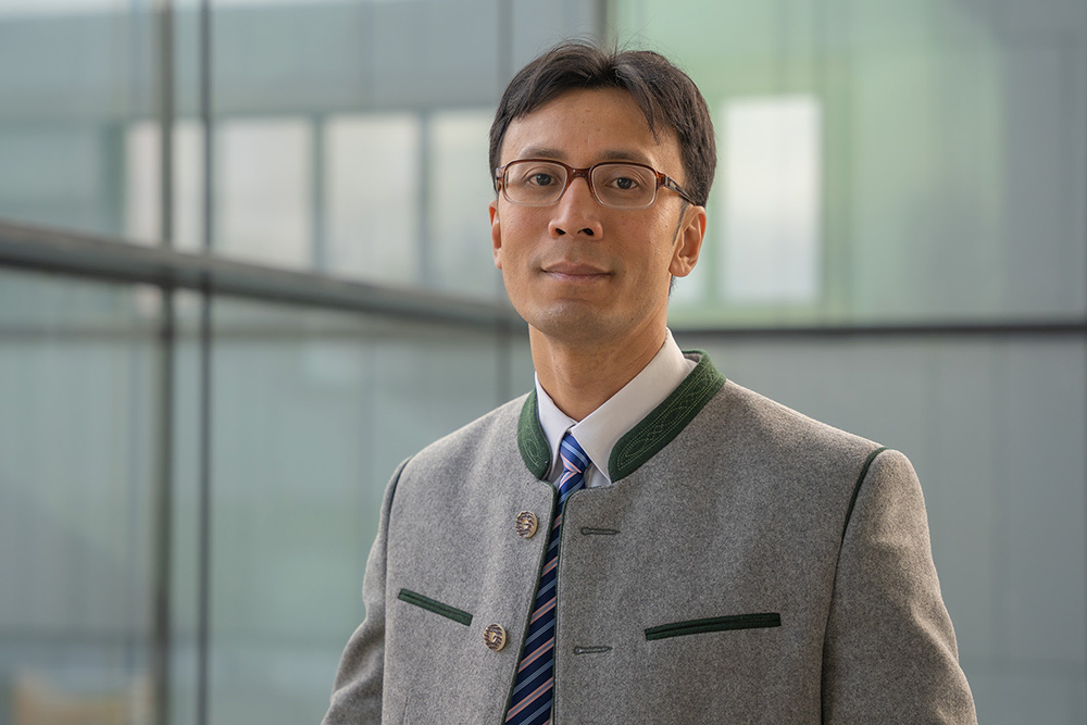
Prof. Dr. med. habil. Thiha Aung, MHBA (Univ.)

Professor

  • Studiengangsleiter Physician Assistant

  • Leiter des Lern- und Transferzentrum/Simulationszentrum

  • Inhaber einer Forschungsprofessur

  • Leiter der Arbeitsgruppe Translationale Sarkomforschung und Lymph-(Angiogenese) Universität Regensburg

LA 27-2.17

0991/3615-8253


Sprechzeiten

nach Vereinbarung


Sortierung:
Zeitschriftenartikel

  • L. Mahl
  • J. Ollig
  • V. Schweihofer
  • L. Wiegand
  • Phillipp Torkler
  • S. Haerteis
  • Thiha Aung

Importance and implications of exosomes in nephrology and urology

In: Pflügers Archiv : European Journal of Physiology

  • 18.11.2022 (2022)

DOI: 10.1007/s00424-022-02771-y

Exosomes are extracellular vesicles that are formed by two invaginations of the plasma membrane and can be released by all eukaryotic cells. Because of their bioactive contents, including nucleic acids and proteins, exosomes can activate a variety of functions in their recipient cells. Due to the plethora of physiological and pathophysiological functions, exosomes have received a lot of attention from researchers over the past few years. However, there is still no consensus regarding isolation and characterization protocols of exosomes and their subtypes. This heterogeneity poses a lot of methodical challenges but also offers new clinical opportunities simultaneously. So far, exosome-based research is still mostly limited to preclinical experiments and early-stage clinical trials since the translation of experimental findings remains difficult. Exosomes could potentially play an important role as future diagnostic and prognostic agents and might also be part of the development of new treatment strategies. Therefore, they have previously been investigated in a variety of nephrological and urological conditions such as acute kidney injury or prostate cancer.
  • Angewandte Informatik
  • Angewandte Gesundheitswissenschaften
  • GESUND
Zeitschriftenartikel

  • E. Pion
  • J. Karnosky
  • S. Boscheck
  • B. Wagner
  • K. Schmidt
  • S. Brunner
  • H. Schlitt
  • Thiha Aung
  • C. Hackl
  • S. Haerteis

3D In Vivo Models for Translational Research on Pancreatic Cancer: The Chorioallantoic Membrane (CAM) Model

In: Cancers vol. 14

  • 31.07.2022 (2022)

DOI: 10.3390/cancers14153733

Pancreatic ductal adenocarcinoma (PDAC) is a highly aggressive cancer with adverse outcomes that have barely improved over the last decade. About half of all patients present with metastasis at the time of diagnosis, and the 5-year overall survival rate across all stages is only 6%. Innovative in vivo research models are necessary to combat this cancer and to discover novel treatment strategies. The chorioallantoic membrane (CAM) model represents one 3D in vivo methodology that has been used in a large number of studies on different cancer types for over a century. This model is based on a membrane formed within fertilized chicken eggs that contain a dense network of blood vessels. Because of its high cost-efficiency, simplicity, and versatility, the CAM model appears to be a highly valuable research tool in the pursuit of gaining more in-depth insights into PDAC. A summary of the current literature on the usage of the CAM model for the investigation of PDAC was conducted and subdivided into angiogenesis, drug testing, modifications, personalized medicine, and further developments. On this comprehensive basis, further research should be conducted on PDAC in order to improve the abysmal prognosis of this malignant disease.
  • Angewandte Gesundheitswissenschaften
  • GESUND
Zeitschriftenartikel

  • E. Pion
  • I. Zucal
  • J. Troebs
  • A.-L. Feder
  • T. Kyaw
  • S. Khin
  • P. Heidekrueger
  • L. Prantl
  • S. Härteis
  • Thiha Aung

New, Innovative, Three-Dimensional In Vivo Model for High-Level Microsurgical and Supermicrosurgical Training: A Replacement for Animal Models

In: Plastic and Reconstructive Surgery (Journal of the American Society of Plastic Surgeons) vol. 150 pg. 432-436.

  • 09.06.2022 (2022)

DOI: 10.1097/PRS.0000000000009330

Microsurgery and supermicrosurgery are surgical subdomains necessary for a large variety of surgical disciplines. So far, there is no training model for lymphatic surgery or perforator flap surgery, and the most commonly used microsurgical training models are living animals. However, the ethical principles of replacement, refinement, and reduction (the three Rs) of living animals for training purposes were implemented, highlighting the necessity of an animal-sparing microsurgical training model. Formed during embryogenesis, the chick chorioallantoic membrane resembles a highly vascularized, noninnervated membrane within fertilized chicken eggs. The aim of this study was to utilize the chorioallantoic membrane model as an innovative and versatile training model for supermicrosurgery and microsurgery that can reduce the number of animals used for these purposes. The variety of different sized vessels for the implementation of an anastomosis proved the chorioallantoic membrane model as a well-functioning supermicrosurgical and microsurgical training model. The circulatory system is resilient enough to withstand the mechanical stress applied to the tissue, and the patency of the implemented anastomosis can be tested for the verification of the procedures. In summary, the integration of the chorioallantoic membrane model into a surgical training program can benefit its quality by representing a realistic anatomical and physiological model with a high variety of vascular structures. Moreover, the chorioallantoic membrane model satisfies the principles of replacement, refinement, and reduction as an animal-sparing model, indicating the potential of this model as an innovative microsurgical training model for the improvement of surgical skills.
  • Angewandte Gesundheitswissenschaften
  • GESUND
Zeitschriftenartikel

  • P. Kuri
  • E. Pion
  • L. Mahl
  • P. Kainz
  • S. Schwarz
  • C. Brochhausen
  • Thiha Aung
  • S. Haerteis

Deep Learning-Based Image Analysis for the Quantification of Tumor-Induced Angiogenesis in the 3D In Vivo Tumor Model-Establishment and Addition to Laser Speckle Contrast Imaging (LSCI)

In: Cells vol. 11

  • 28.07.2022 (2022)

DOI: 10.3390/cells11152321

(1) Background: angiogenesis plays an important role in the growth and metastasis of tumors. We established the CAM assay application, an image analysis software of the IKOSA platform by KML Vision, for the quantification of blood vessels with the in ovo chorioallantoic membrane (CAM) model. We added this proprietary deep learning algorithm to the already established laser speckle contrast imaging (LSCI). (2) Methods: angiosarcoma cell line tumors were grafted onto the CAM. Angiogenesis was measured at the beginning and at the end of tumor growth with both measurement methods. The CAM assay application was trained to enable the recognition of in ovo CAM vessels. Histological stains of the tissue were performed and gluconate, an anti-angiogenic substance, was applied to the tumors. (3) Results: the angiosarcoma cells formed tumors on the CAM that appeared to stay vital and proliferated. An increase in perfusion was observed using both methods. The CAM assay application was successfully established in the in ovo CAM model and anti-angiogenic effects of gluconate were observed. (4) Conclusions: the CAM assay application appears to be a useful method for the quantification of angiogenesis in the CAM model and gluconate could be a potential treatment of angiosarcomas. Both aspects should be evaluated in further research.
  • Angewandte Gesundheitswissenschaften
  • GESUND
Zeitschriftenartikel

  • C. Kohl
  • Thiha Aung
  • S. Haerteis
  • A. Ignatov
  • O. Ortmann
  • T. Papathemelis

The 3D in vivo chorioallantoic membrane model and its role in breast cancer research

In: Journal of Cancer Research and Clinical Oncology vol. 148 pg. 1033-1043.

  • 05.02.2022 (2022)

DOI: 10.1007/s00432-022-03936-z

PURPOSE We aimed to evaluate the role of the chorioallantoic membrane model (CAM) in breast cancer research. METHODS The following is an overview of the use of the CAM in the field of breast cancer research based on a PubMed literature query. RESULTS The CAM is a 3D in vivo model that can be used for the analysis of tumor growth, biology and angiogenesis of primary tumor tissue or tumor cell lines. The CAM model has been used in breast cancer research for drug testing, migration assays and the evaluation of vascularization, amongst others. The CAM model is a valuable method that offers a better imitation of the physiological phenomena compared to 2D or 3D in vitro models. CONCLUSION The CAM model has primarily and successfully been utilized for the assessment of the tumor biology of established breast cancer cell lines. Further, the CAM model is a promising method to analyze patient derived primary tumor material and could be used as a "patient-specific 3D-tumor-therapy-model" for the cost-efficient evaluation of anti-cancer drugs to find the optimal treatment for breast cancer patients.
  • Angewandte Gesundheitswissenschaften
  • GESUND
Zeitschriftenartikel

  • E.-M. Bichlmayer
  • L. Mahl
  • Leo Hesse
  • Eric Pion
  • Victoria Haller
  • Andreas Moehwald
  • Christina Hackl
  • Jens Werner
  • Hans Schlitt
  • Siegfried Schwarz
  • Philipp Kainz
  • Christoph Brochhausen
  • Christian Groeger
  • Felix Steger
  • Oliver Kölbl
  • Christoph Daniel
  • Kerstin Amann
  • Andre Kraus
  • Björn Buchholz
  • Thiha Aung
  • Silke Haerteis

A 3D In Vivo Model for Studying Human Renal Cystic Tissue and Mouse Kidney Slices

In: Cells vol. 11

  • 22.07.2022 (2022)

DOI: 10.3390/cells11152269

(1) Background: Autosomal dominant polycystic kidney disease (ADPKD) is a frequent monogenic disorder that leads to progressive renal cyst growth and renal failure. Strategies to inhibit cyst growth in non-human cyst models have often failed in clinical trials. There is a significant need for models that enable studies of human cyst growth and drug trials. (2) Methods: Renal tissue from ADPKD patients who received a nephrectomy as well as adult mouse kidney slices were cultured on a chorioallantoic membrane (CAM) for one week. The cyst volume was monitored by microscopic and CT-based applications. The weight and angiogenesis were quantified. Morphometric and histological analyses were performed after the removal of the tissues from the CAM. (3) Results: The mouse and human renal tissue mostly remained vital for about one week on the CAM. The growth of cystic tissue was evaluated using microscopic and CT-based volume measurements, which correlated with weight and an increase in angiogenesis, and was accompanied by cyst cell proliferation. (4) Conclusions: The CAM model might bridge the gap between animal studies and clinical trials of human cyst growth, and provide a drug-testing platform for the inhibition of cyst enlargement. Real-time analyses of mouse kidney tissue may provide insights into renal physiology and reduce the need for animal experiments.
  • Angewandte Gesundheitswissenschaften
  • GESUND
Zeitschriftenartikel

  • K. Drexler
  • B. Schwertner
  • S. Haerteis
  • Thiha Aung
  • M. Berneburg
  • E. Geissler
  • M. Mycielska
  • S. Haferkamp

The Role of Citrate Homeostasis in Merkel Cell Carcinoma Pathogenesis

In: Cancers vol. 14

  • 14.07.2022 (2022)

DOI: 10.3390/cancers14143425

Merkel cell carcinoma (MCC) is a rare but highly aggressive tumor of the skin with a poor prognosis. The factors driving this cancer must be better understood in order to discover novel targets for more effective therapies. In the search for targets, we followed our interest in citrate as a central and critical metabolite linked to fatty acid synthesis in cancer development. A key to citrate uptake in cancer cells is the high expression of the plasma membrane citrate transporter (pmCiC), which is upregulated in the different adenocarcinoma types tested so far. In this study, we show that the pmCiC is also highly expressed in Merkel cell carcinoma cell lines by western blot and human tissues by immunohistochemistry staining. In the presence of extracellular citrate, MCC cells show an increased proliferation rate in vitro; a specific pmCiC inhibitor (Na+-gluconate) blocks this citrate-induced proliferation. Furthermore, the 3D in vivo Chick Chorioallantoic Membrane (CAM) model showed that the application of Na+-gluconate also decreases Merkel cell carcinoma growth. Based on our results, we conclude that pmCiC and extracellular citrate uptake should be considered further as a potential novel target for the treatment of Merkel cell carcinoma.
  • Angewandte Gesundheitswissenschaften
  • GESUND
Zeitschriftenartikel

  • L. Sommerauer
  • A. Phyo
  • E. Pion
  • I. Zucal
  • E. Klingelhoefer
  • S. Thu
  • T. Win
  • S. Khin
  • T. Kyaw
  • H. Zaw
  • M. Htwe
  • N. Fabbri
  • S. Haerteis
  • Thiha Aung

Modified Borggreve-Van Nes-Winkelmann rotationplasty for surgery in developing countries

In: BMC Surgery vol. 22 pg. 333.

  • 07.09.2022 (2022)

DOI: 10.1186/s12893-022-01780-z

BACKGROUND Amputation is still the most common therapy for patients suffering from osteosarcoma in Myanmar, despite the fact that limb salvage surgery e.g. Borggreve-Van Nes-Winkelmann rotationplasty for malignant tumors located within the distal femur or proximal tibia is the current state-of-the-art reconstructive procedure. A safe and reliable operation technique is crucial in order to perform a complex surgical procedure like the rotationplasty in lower-middle income economies with limited infrastructure and resources. The authors present seven cases of patients with osteosarcomas that received a Borggreve-Van Nes-Winkelmann rotationplasty with an evaluation of the procedures focusing on safety and sustainability. METHODS From 2019 until 2020, seven young patients with osteosarcomas of the distal femur or proximal tibia were treated with Borggreve-Van Nes-Winkelmann rotationplasties in the Orthopaedic Hospital in Mandalay, Myanmar. As modification of the standard procedure the dissection and subsequent clamping of the femoral artery in order to minimize blood loss as well as the formation of an adipocutaneous flap that minimizes swelling and decreases the pressure on the vessels were successfully performed. This modified procedure resembles a safe and simplified surgical technique that is feasible under the circumstances of lower-middle income economies with good outcomes. RESULTS All patients showed good functional and aesthetic results. One of the seven patients needed secondary wound closure due to wound dehiscence. CONCLUSIONS A simplified and safe operation technique for the performance of the Van Nes-Borggreve rotationplasty was adapted to the given constraints in lower-middle income economies and proved to be successful. Trial registration All patients approved to participate in the study and have given consent to publication.
  • Angewandte Gesundheitswissenschaften
  • GESUND
Zeitschriftenartikel

  • Isabel Zucal
  • Anna-Lena Feder
  • Thura Kyaw
  • Sopyay Khin
  • Paul Heidekrueger
  • Lukas Prantl
  • Silke Haerteis
  • Thiha Aung

An Innovative Simulation Model for Microvascular Training

In: Plastic and Reconstructive Surgery (Journal of the American Society of Plastic Surgeons) vol. 150 pg. 189e-193e.

  • 29.06.2022 (2022)

DOI: 10.1097/PRS.0000000000009209

Preclinical/clinical microsurgical training is essential for clinical practice. Therefore, various training models have been established, such as synthetic and cadaveric models. The most common limitation of these models is the lack of circulation, which limits the simulation of real intraoperative circumstances. Thus, the authors aimed to create a novel model that provides blood circulation with an extracorporeal perfusion device that they attached to rat cadavers for the reestablishment of a circulatory system. Patent blue and heparin were added to the perfusion fluid to visualize circulation and to dissolve thrombosis, and indocyanine green fluorescent imaging was applied to show the perfusion of the entire body. The femoral and brachial vessels were dissected, and an end-to-end anastomosis was performed on the femoral artery. The patency of the operated vessel was visualized with indocyanine green fluorescent imaging. Indocyanine green fluorescent imaging showed appropriate vessel patency and extremity perfusion through the anastomosis. The use of this novel rat model enables a solution for ethical problems encountered when using rats for surgical training courses. By practicing on these animal-sparing models with intact circulation, microsurgical skills can be improved. Future studies on further microsurgical techniques and vascular perfusion of organs or tumors may benefit from our model.
  • Angewandte Gesundheitswissenschaften
  • GESUND
Zeitschriftenartikel

  • I. Zucal
  • S. Geis
  • L. Prantl
  • S. Haerteis
  • Thiha Aung

Indocyanine Green for Leakage Control in Isolated Limb Perfusion

In: Journal of Personalized Medicine vol. 11

  • 05.11.2021 (2021)

DOI: 10.3390/jpm11111152

Sarcomas are characterized by a high metastatic potential and aggressive growth. Despite surgery, chemotherapy plays an important role in the treatment of these tumors. Optimal anti-cancer therapy with maximized local efficacy and minimized systemic side effects has been the object of many studies for a long time. To improve the local efficacy of anti-tumor therapy, isolated limb perfusion with high-dose cytostatic agents has been introduced in surgical oncology. In order to control the local distribution of substances, radiolabeled cytostatic drugs or perfusion solutions have been applied but often require the presence of specialized personnel and result in a certain exposure to radiation. In this study, we present a novel strategy using indocyanine green to track tumor perfusion with high-dose cytostatic therapy. In a rat cadaver model, the femoral vessels were cannulated and connected to a peristaltic pump to provide circulation within the selected limb. The perfusion solution contained indocyanine green and high-dose doxorubicin. An infrared camera enabled the visualization of indocyanine green during limb perfusion, and subsequent leakage control was successfully performed. Histologic analysis of sections derived proximally from the injection site excluded systemic drug dispersion. In this study, the application of indocyanine green was proven to be a safe and cost- and time-efficient method for precise leakage control in isolated limb perfusion with a high-dose cytostatic agent.
  • Angewandte Gesundheitswissenschaften
  • GESUND

Projekte

* AI Augmented Decision Support System **Personalisierte Medizin in der Onkologie und Nephrologie unter Einsatz von 3D-in-vivo-Modellen


Labore

*Lern- und Transferzentrum/Simulationszentrum **Lehrstuhl für Molekulare und Zelluläre Anatomie, Universität Regensburg (S1/S2 Laboren)


Kernkompetenzen

  • Facharzt für Plastische und Ästhetische Chirurgie
  • Plastisch-Rekonstruktiver Tumorchirurg
  • Mikro- und Supermikrochirugie
  • Lymphchirurgie


Forschungs- und Lehrgebiete

  • AI Augmented Decision Support System

  • Spezielle Visualisierungstechniken in der Lehre mit 3D-Anwendungen - Schwerpunkt auf virtuelle Präparationen in der Anatomie

  • Visualisierung und KI im Bereich der Angiogenese

  • Rehabilitation nach onkologischen und traumatischen Amputationen, Moderne Prothesenanpassung, Targeted-Muscle-Reinnervation (TMR) Versorgung von Prothesen

  • Translationale onkologische Forschung

  • Leiter der Arbeitsgruppe Translationale Sarkomforschung und Lymph-(Angiogenese) - siehe: https://www.uni-regensburg.de/biologie-vorklinische-medizin/molekulare-zellulaere-anatomie-haerteis/mitarbeiter/index.html

Forschung gefördert durch:

  • Deutsche Forschungsgemeinschaft Sonderforschungsbereich SFB 1350 / Transregio TRR 374
  • Deutsche Forschungsgemeinschaft (DFG)
  • Bundesministerium für Bildung und Forschung (BMBF)
  • Else-Kröner-Fresenius Stiftung
  • Deutsche Gesellschaft für Internationale Zusammenarbeit (GIZ) GmbH
  • „Zentrales Innovationsprogramm Mittelstand“ des Bundesministeriums für Wirtschaft und Energie (BMWi; Projektform FuE-Kooperationsprojekte; AiF Projekt GmbH)

Bei Interesse an Bachelor-/Master-/PhD arbeiten und Medizinischen Doktorarbeiten bitte per Email anfragen.
Wir bieten:

  • Kombination aus experimenteller Forschung und klinischem Mentoring

  • Internationale Forschungsaufenthalt in Kooperation mit Harvard Medical School

  • Internationale Hospitationen und Mitwirkung bei humanitären medizinischen Hilfsprojekten

  • https://www.uni-regensburg.de/biologie-vorklinische-medizin/molekulare-zellulaere-anatomie-haerteis-1/publikationen/index.html


Vita

  • Humanmedizin-Studium an der Universität Göttingen
  • Promotion zum Dr. med. an der Universität Göttingen
  • Habilitation zum Dr. med. habil. an der Universität Regensburg
  • Facharzt für Plastische und Ästhetische Chirurgie
  • Plastisch-Rekonstruktiver Tumorchirurg
  • MHBA an der Friedrich-Alexander-Universität Erlangen-Nürnberg


Sonstiges

Auslandsaufenthalte:

  • Hospital de la Santa Creu i Sant Pau, Abteilung Plastische Chirurgie, Schwerpunkt Lymphchirurgie, Barcelona (Spanien)

  • Universitätsklinik Gent (Belgien), Department of Plastic Surgery of the University Hospital Gent - Schwerpunkt Lymphchirurgie

  • Universitätsklinik Tokyo (Japan), Abteilung Plastische Chirurgie, Schwerpunkt Rekonstruktion, Replantation und Lymphchirurgie

  • Linkou Chang Gung Memorial Hospital, Taoyan (Taiwan), Division of Plastic Reconstructive Microsurgery

  • Memorial-Sloan-Kettering Cancer Center, Department of Surgery, Orthopaedic Service, New York, USA

  • Mayo Clinic, Division of Plastic Surgery, Mayo Clinic, Jacksonville, Florida,USA

  • Harvard Medical School, Beth Israel Deaconess Medical Center, Division of Plastic and Reconstructive
    Surgery, Boston, Massachusetts, USA

Publikationen: https://www.uni-regensburg.de/biologie-vorklinische-medizin/molekulare-zellulaere-anatomie-haerteis-1/publikationen/index.html